The global pharmaceutical industry operates under a costly paradox: nearly 95% of drug candidates validated in animal models ultimately fail in human clinical trials. This translation gap drains billions of dollars annually in wasted R&D expenditure and delays life-saving therapeutics from reaching patients.
In response, global regulatory authorities are enacting an unprecedented paradigm shift:
- The U.S. FDA Modernization Act 2.0 formally eliminated the mandate for animal testing in preclinical drug development, opening the door for New Approach Methodologies (NAMs).
- The FDA’s formal draft guidance on NAM validation principles signals a transition from regulatory permission to explicit regulatory expectation.
- Programs such as the NIH CARE initiative ($150 million) and the Organoid Research Alliance ($87 million) underscore that 3D human-relevant models are becoming the new baseline for preclinical safety and efficacy profiling.
However, as highlighted in a strategic report by market intelligence firm Alcimed (“Organoids for drug development: 3 strategic considerations for your preclinical pipeline”, 2026), widespread industrial adoption of organoids remains severely constrained by a lack of standardization and high assay variability.
Why standard 3D models fail regulatory & industrial standards
While stem-cell-derived organoids offer superior biological relevance compared to 2D cell cultures, most drug discovery programs still grow them in undefined animal-derived matrices (e.g., Matrigel) or basic synthetic hydrogels.
This creates three critical engineering bottlenecks that undermine clinical predictability:
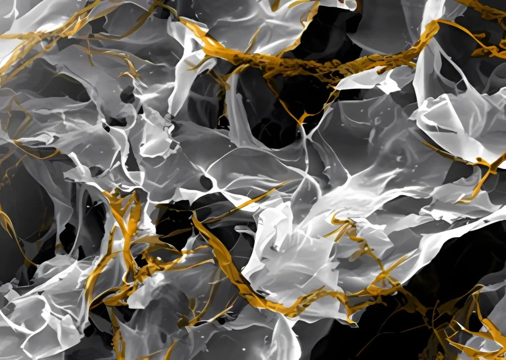
- Uncontrolled lot-tolot variability: Complex animal-derived substrates contain ill-defined growth factors and batch-dependent compositions, making reproducible high-throughput screening (HTS) virtually impossible across multi-site studies.
- Mechanical misalignment with human tissues: Passive hydrogels fail to replicate the dynamic biomechanical microenvironment of human organ tissue—such as stiffness variations between healthy organ stroma, fibrotic tissue, and solid tumor microenvironments.
- Artifacts in toxicity & efficacy data: Cells cultured without proper biomechanical cues display altered gene expression and aberrant signaling pathways, leading to false positives and misleading toxicological readouts early in the pipeline.
In short: culturing sophisticated human organoids inside uncharacterized, non-physiological supports compromises data integrity at the most critical stage of drug discovery.
The biomimetic engine: physioscaffolds as the benchmark for reproducibility
To meet the rigorous validation standards set by the FDA and global health agencies, 3D cell culture must evolve from empirical biology to precision microenvironment engineering.
This is the core paradigm behind Organisyl’s Physioscaffold architecture.
Rather than relying on uncharacterized biological matrices or low-end passive hydrogels, Organisyl utilizes a fully defined, synthetic extracellular matrix engineered from grafted hyaluronic acid (HAphy).
This platform directly resolves the technical limitations identified by industry analysts:
- Zero lot-to-lot variation: Being 100% chemically defined, the HAphy Physioscaffold guarantees identical physicochemical properties across 96-well plates, fulfilling the strict reproducibility requirements of regulatory audits and automated HTS workflows.
- Tunable biomechanical fidelity (100 Pa to 20 kPa): The Physioscaffold allows R&D teams to tune matrix stiffness and viscoelasticity to match the exact physiological signature of target human tissues—from soft neural networks to rigid tumor stromas.
- Restored phenotypic relevance: By embedding organoids and primary cells within their native biomechanical niche, the Physioscaffold preserves physiological cell-matrix signaling, significantly improving predictive accuracy for Drug-Induced Liver Injury (DILI) and target engagement.
Strategic roadmap: de-risking your transition to NAMs
As regulatory expectations tighten, delaying the adoption of standardized 3D microenvironments poses a direct risk to pipeline valuation. Early clinical attrition can be systematically mitigated by optimizing preclinical culture supports.
For R&D leaders and Preclinical Safety heads, we recommend a three-step evaluation process:
- Audit current 3D matrix protocols: Pinpoint where lot-to-lot matrix variability introduces statistical noise in your lead optimization phase.
- Standardize the microenvironment: Replace undefined animal matrices with chemically defined Physioscaffolds to establish robust, audit-ready data sets for regulatory submissions.
- Integrate tissue biomechanics: Account for tissue stiffness in safety and efficacy screens to eliminate toxic candidates before capital-intensive IND-enabling studies.
Ready to de-risk your preclinical pipeline?
Whether you are transitioning toward FDA-compliant NAMs or looking to eliminate batch-to-batch noise in automated HTS, our team is here to help => [Schedule a strategic pipeline review]